Vision depends on several delicate structures working together, and the retina plays one of the most important roles. Located at the back of the eye, the retina receives light and converts it into signals that are sent to the brain. When the retina separates from its normal position, a condition known as retinal detachment can develop. Because prolonged separation can damage retinal tissue, prompt medical assessment is extremely important.
For people searching for retinal detachment surgery in ghaziabad, understanding the symptoms, causes, available procedures, and recovery process can help them respond appropriately when vision changes occur. Choosing a well-equipped Eye hospital in Ghaziabad is also important because retinal conditions often require detailed examination, specialized equipment, and timely intervention.
What Is Retinal Detachment?
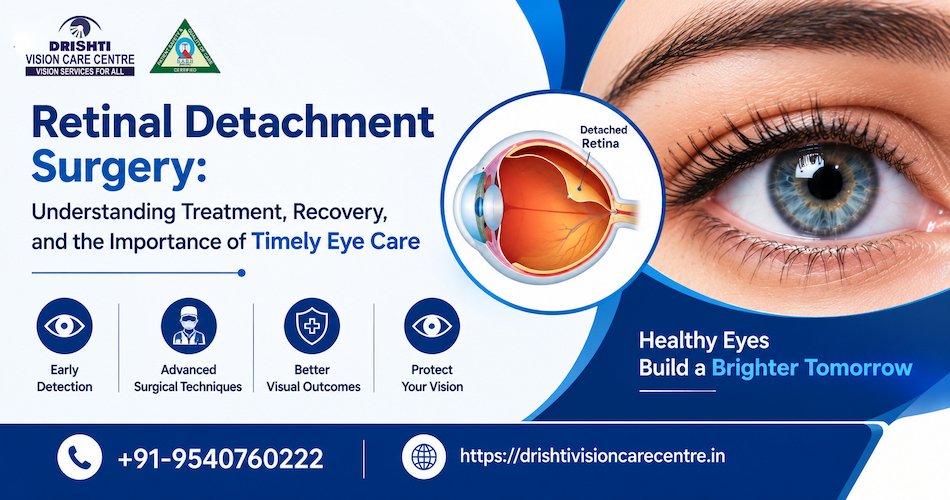
Retinal detachment occurs when the retina becomes separated from the tissues that support it. This prevents the retina from functioning normally and can affect vision significantly.
A retinal detachment may develop because of a retinal tear, changes in the vitreous gel inside the eye, an eye injury, previous eye surgery, or certain retinal conditions. In some cases, it can progress quickly and become a sight-threatening emergency.
The condition is different from ordinary changes in eyesight and should not be ignored.
Recognizing the Early Warning Signs
One of the most important aspects of retinal health is recognizing symptoms early. Retinal detachment may not initially cause pain, which can sometimes lead people to delay an examination.
Potential warning signs include:
- A sudden increase in eye floaters
- Flashes of light in the field of vision
- A dark shadow or curtain-like area
- Sudden reduction in vision
- Distortion or changes in peripheral vision
- A noticeable change in one eye compared with the other
Experiencing these symptoms does not necessarily mean that retinal detachment has occurred, but they require prompt assessment by an eye specialist.
Why Prompt Treatment Matters
The retina needs an adequate blood supply and proper contact with the underlying tissues to function normally. When it becomes detached, prolonged separation can compromise retinal function.
Early diagnosis can allow the ophthalmologist to determine whether a retinal tear, partial detachment, or more extensive separation is present. Depending on the findings, treatment may range from laser procedures for certain retinal tears to surgery for an established detachment.
This is why people looking for an Eye hospital in Ghaziabad should consider facilities that can provide comprehensive retinal evaluation rather than delaying care when symptoms appear.
How Is Retinal Detachment Diagnosed?
Diagnosis begins with a detailed eye examination. The ophthalmologist may dilate the pupil to examine the retina and identify tears, holes, fluid accumulation, or areas of detachment.
Additional diagnostic techniques may include:
Dilated Retinal Examination
A dilated examination allows the doctor to view a larger portion of the retina and assess its condition.
Optical Coherence Tomography
OCT imaging can provide detailed cross-sectional views of retinal structures and may be useful when assessing associated retinal changes.
Ocular Ultrasound
When the view of the retina is limited, ultrasound imaging can help the ophthalmologist evaluate the structures inside the eye.
The combination of clinical examination and appropriate imaging helps determine the most suitable treatment.
When Is Retinal Detachment Surgery Required?
Not every retinal problem requires the same procedure. Treatment depends on the location and size of the detachment, the presence of retinal tears, the condition of the vitreous, and whether the central retina is involved.
Surgery may be recommended when the retina has become detached and needs to be repositioned and secured.
Common surgical approaches include pneumatic retinopexy, scleral buckling, and vitrectomy. In some cases, a combination of techniques may be used.
The ophthalmologist selects the procedure based on the individual retinal condition rather than applying one approach to every patient.
Understanding Vitrectomy
Vitrectomy is a retinal surgery in which some or all of the vitreous gel is removed to provide access to the retina. The surgeon can then address retinal tears or other abnormalities and use a suitable method to support retinal reattachment.
A temporary gas bubble or silicone oil may sometimes be used to help keep the retina in position while it heals.
Patients receiving a gas bubble may receive specific instructions regarding head positioning and air travel. These instructions are important and should be followed carefully.
What Happens During Scleral Buckling?
Scleral buckling is another surgical technique used for selected retinal detachments. A flexible band is placed around the outside of the eye to provide support to the weakened area and help relieve traction on the retina.
The exact approach depends on the characteristics of the retinal detachment. The ophthalmologist determines whether this procedure, vitrectomy, or another technique is appropriate.
Preparing for Retinal Surgery
Before surgery, patients generally undergo a detailed assessment of their eye and overall health. The ophthalmologist explains the recommended procedure, potential risks, expected recovery, and postoperative requirements.
Patients should provide accurate information about medications, previous surgeries, allergies, and existing health conditions.
Understanding the instructions before surgery can make the treatment process easier and help patients prepare for the recovery period.
Recovery After Retinal Detachment Surgery
Recovery varies depending on the surgical technique and severity of the retinal condition. Vision may remain blurred for some time after surgery, particularly when a gas bubble or other internal support is used.
During recovery, patients may be advised to:
- Use prescribed eye drops correctly
- Avoid rubbing or pressing on the eye
- Follow activity restrictions
- Maintain the recommended head position when instructed
- Attend scheduled follow-up appointments
- Report unusual pain, worsening redness, or sudden vision changes promptly
Follow-up examinations allow the ophthalmologist to monitor retinal healing and identify complications at an early stage.
Choosing the Right Eye Care Centre
Retinal surgery requires specialized knowledge, diagnostic capabilities, and careful postoperative monitoring. When evaluating an Eye hospital in Ghaziabad, patients should consider the availability of retinal specialists, diagnostic facilities, surgical infrastructure, and follow-up care.
It is also important to select a centre where treatment recommendations are based on detailed examination rather than assumptions about the condition.
Retinal Care at Drishti Vision Care Centre
Drishti Vision Care Centre provides comprehensive ophthalmic services with an emphasis on detailed diagnosis and individualized treatment planning. For patients experiencing symptoms that may indicate a retinal problem, timely evaluation can help determine the condition and appropriate next steps.
Retinal assessment may involve clinical examination and advanced diagnostic imaging when required. Treatment recommendations are based on the patient’s retinal findings, visual condition, and overall eye health.
For individuals considering retinal detachment surgery in ghaziabad, an early consultation can be an important step toward understanding the condition and available treatment options.
Can Retinal Detachment Be Prevented?
Not every retinal detachment can be prevented, but regular eye examinations can help identify certain risk factors and retinal tears before they progress.
People with significant short-sightedness, previous eye surgery, eye trauma, a history of retinal problems, or a family history of retinal detachment may benefit from regular retinal evaluations.
Most importantly, sudden flashes, new floaters, or a curtain-like shadow should not be treated as an ordinary vision problem.
Retinal detachment is a serious condition in which timely diagnosis and appropriate treatment can be critical for preserving vision. Recognizing warning signs and seeking an immediate eye examination can help identify retinal problems before they progress further.
If you are searching for retinal detachment surgery hospital in ghaziabad, discuss your symptoms with a qualified retinal specialist and undergo a comprehensive evaluation. A well-equipped Eye hospital in Ghaziabad can provide the diagnostic assessment and treatment guidance needed for complex retinal conditions.
With specialized eye care, appropriate surgical planning, and consistent follow-up, patients can receive personalized management based on their individual retinal health. Drishti Vision Care Centre focuses on comprehensive evaluation and patient-oriented eye care to help individuals make informed decisions about their vision.